|
|
|
MODEL Version
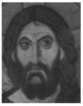
Static eye Operation of the ModelTo start using this model, input any gray scale 8bit bitmapped image. The size is limited by the power of your computer and your patience in waiting for output. Here is an example Image:
Next the parameters of the model are set. The only parameters to set are the dimensions of the excitatory and inhibitory fields:
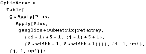
se and si substitute for se and si and width is in place of w in the equation before. The ganglion cell is defined in Mathematica® as shown below:
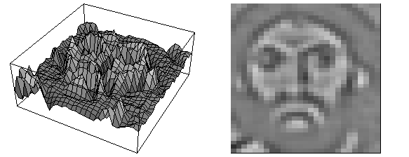
The ganglion cell is then point by point multiplied with a submatrix of the same size as shown below. The is done repeatedly with the center of the ganglion cell being moved in regular steps over the surface (in this case in steps of five pixels). The code is shown first and then the output is plotted so that the stronger the output the more light the graph. Q, below, is a constant that is used to scale the output. One of the features of the scale is to keep all of the outputs about equal in range. If the standard deviations of the excitatory and inhibitory regions increase, the range of outputs generally increase as a result of greater numbers in the sum (seen in the two apply steps in the code). The Q is partly scaled by the size of the ganglion cell array to compensate for that fact.
This receptive field is fairly small and you can see that it is very sensitive to edge and fine details. Here is a page devoted to how the model reveals the Hermann Grid illusion.
|